Order Entry
ContactUsLinkComponent

Histology

When studying cells and tissues, and there are various techniques and methods for preparing specimens for examination.

For microscopic study, staining is used to highlight important features of the tissue, but no single inspection technique will display all the cells of a tissue equally. More commonly, sections of a tissue sample are prepared to be examined by several types of microscopes.

That’s why choosing the right histology equipment and supplies is so important. Avantor® can help by offering everything you need to conduct your cutting-edge histological research and medical tests. 

260x190-Recovered-Recovered.jpg

Avantor is committed to providing histology labs with a start-to-finish portfolio of histology products, enabling you to achieve your goals.

Contact us today to discuss your needs. Our team is ready to help.

Contact us

Equipment & instruments

Fume hoods provide the necessary protection for both the working materials and personnel.

Prevent wrinkling and distortion of your paraffin-embedded tissue sections one of our offers.

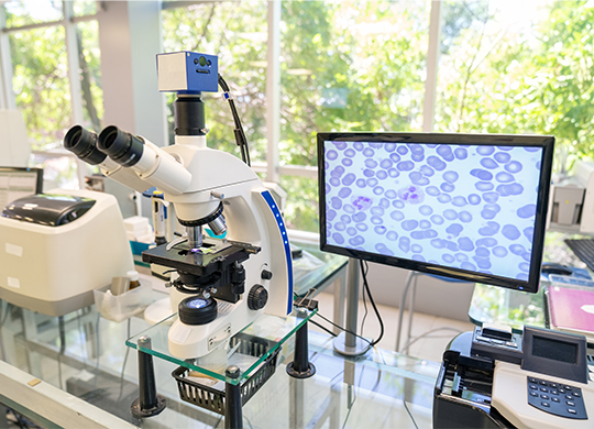
Our catalog of microscopes contain many options for sample analysis and evaluation and use on many applications. Some varieties shine additional illumination directly through the sample to throw images into sharp relief, while others rely on reflected light to highlight surface details in samples that are too thick or opaque for conventional backlighting to be effective.

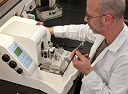
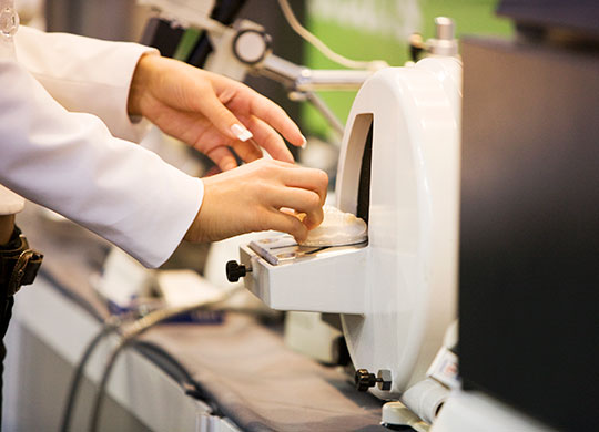
Microtomes use steel, glass, or diamond blades, based on the specimen and hardness, to cut very thin sample slices needed for routine microscopic examinations. Cooled versions are also available.

Heat slides and maintain optimal temperature for optimal adhesion of tissue sections during staining procedures.

Embed tissue samples in a solid medium for thin sectioning and histological analysis.

Reagents and processors to prepare tissues safely and effectively for histological study.

Paraffin wax trimmers remove excess paraffin from embedding tissue cassettes. Available features include high and low temperature settings and magnetic collection trays.

Reagents

Extensive offering of paraffin for histology applications.

A broad line of reagent droppers, diagnostic kits, antigens and antisera to identify a variety of microscopic organisms.

Consumables

Unlike scalpels, biopsy punches manually extract full-thickness skin specimens in diagnostic applications while still maintaining the best cosmetic appearance.

Options feature maximum clarity, superior corrosion resistance and uniform thickness and size.

A wide offering to quickly produce clean slides that will prevent cell and trapped air cross-contamination.

Sets contain instructions and dissecting tools, including scalpels, scissors, forceps, teasing needles and pipettes.

Available in a variety of designs and lengths for specific laboratory procedures.

Microtome blades are used for paraffin and cryostat sectioning.

An extensive offering suited for any application in compounding, including syringe and needle combinations.

A wide range of scalpels for professional use.

Edges are available in both sharp and blunt. Tips come curved, bent or straight.

Numerous options to safely hold tissue specimens submerged in liquid solution for processing.

Embedding Tools
540x190_image.jpg

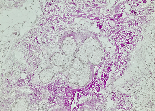
The technique of enclosing tissues or specimens in a mass of embedding media using a mold is known as embedding. During embedding, substances in liquid state are infiltrated through the sample and, after a period of cooling, the embedding substance, and therefore the sample, gets solid without altering the molecular features of tissues.

In addition, embedding is a good method for preserving samples during long periods of time. Several embedding substances are available to get specific section thickness and for performing techniques. For light microscopy studies, paraffin is the most common embedding substance. For electron microscopy, epoxy and acrylic resins are the most used embedding medium. There are other embedding substances, such as celloidin, nitrocellulose, polyethylene glycol and wax polyester.

View our vast range of premium freezer section compounds, dyes and media complemented with the most up-to-date tools, such as Microtome blades, for the process.

Stains
540x190_image_2.jpg

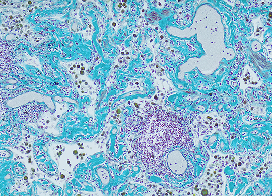
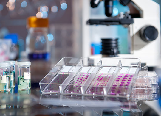
Staining is a technique that can be used to better visualize cells and cell components under a microscope. By using different stains, one can preferentially stain certain cell components, such as a nucleus or a cell wall, or the entire cell.

The most basic reason that cells are stained is to enhance visualization of the cell or certain cellular components under a microscope. Cells may also be stained to highlight metabolic processes or to differentiate between live and dead cells in a sample.

There are many different stains and staining procedures used in histology. Some involve a single stain and just a few steps, while others use multiple stains and a more complicated procedure.

Avantor offers a selection of hematology stains, histology stains, and microbiology stains that are manufactured in accordance with standard specifications to ensure a high level of quality.

Specimen Collection Tools
540x190_image_3.jpg

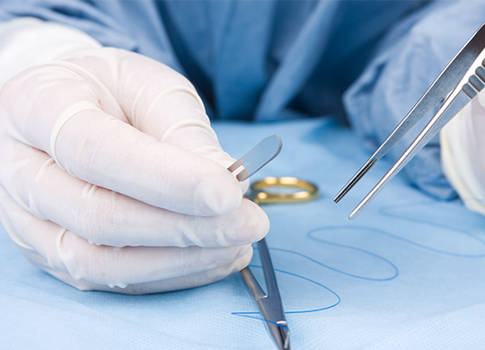
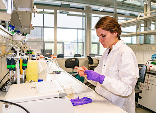
Effectively analyzing a material begins with using the right tools to collect a sample, whether it’s from the body to aid in medical diagnosis, from the environment to identify potentially harmful pathogens, or from a crime scene to aid in an investigation. Various tools, available from Avantor, are required for a proper specimen collection in histology.

  • Scissors - Used for cutting tissue, suture, or for dissection. Scissors can be straight or curved, and may be used for cutting heavy or finer structures.
  • Forceps - Used for grasping tissue or objects. Can be toothed (serrated) or non-toothed at the tip.
  • Needles - Needles come in many shapes and cutting edges for blood collection, biopsies and various other lab applications.
  • Scalpel - Used for initial incision and cutting tissue. Consists of a blade and a handle. Surgeons often refer to the instrument by its blade number.

You can choose tools based on your demands from the variety of materials we offer. Avantor has economical, disposable choices or reusable types for repeated procedural use.

What are the applications of histology?
Accordian Block540x390.jpg

Histology is a versatile scientific study examining the microanatomy of cells and tissues. The sectioning and staining of the tissues can provide vivid imagery of samples, making it possible for researchers to understand the biological processes of a tissue or cell. Applications of histology include: 

Aid in disease diagnosis 
Medical innovations and discovery 
Education and research (ex: Archaeology) 
Forensic analysis In all these fields, having quick and dependable access to consumables is needed to prepare samples and run a histology report effectively. In many cases, laboratory protocols will call for a large quantity of samples to be tested making it critical to optimize your methods to save time and resources.

Two Ways to Optimize Histology Sample Preparation with Lab Consumables
histology-experiment-177713092-540x390.jpg

Proper handling of your samples and laboratory supplies can ensure readable and consistent results. Dependable protocols can take considerable time and effort to establish, making it easy to waste consumable materials and sample tissue in the process. There are ways you can prepare your workspace and team to minimize errors and waste when operating a histology lab.

1. Handle histology samples with care
When it comes to successful histology testing, the correct handling of your sample reduces waste and supports viable results. Cytology samples can be significantly easier to work with if you try the following:

Use microscope slides with frosted ends Rapidly drying your slides after preparation results in superior cell preservation Handling tissue samples in separate containers will prevent contamination or wasted materials. Fix tissues by using the correct ratio of formalin and sample tissue (10:1 by volume).

2. Properly prepare and embed your sample tissue
Before sectioning your sample in the microtome, the tissue sample must be embedded. This is done by immersing the sample in molten paraffin wax or frozen in a solution. This process is key for preserving biological materials in the sample and will aid in preserving both tissue morphology and providing a clean cut during the sectioning phase.

Once a sample is set, use caution when opening lids, as tissue often sticks to them. In cases where tools need to be used, it is good practice to keep your equipment sanitized to avoid any contamination at this phase. Beyond the handling of sample material, selecting the correct histology consumables will provide a foundation for successful bioprocessing. This starts with choosing the right containers and specifically, cassettes that are compatible with your sample materials.

The Difference Between Biopsy Cassettes and Tissue Cassettes
scientist-slide-1214913572-540x390.jpg

Cassettes are essential for the processing of tissue blocks in a histology lab. Both biopsy and tissue cassettes provide a similar mechanism for histology testing. They are made up of materials resistant to histological solvents and are both designed with slots to offer maximum flow through. This enables good fluid exchange and proper drainage from the cassette. The key difference between these two histology lab consumables is the design of the slots in the cassettes.

Biopsy cassettes feature a small grid of circular slots, whereas tissue cassettes have longer slot openings within the tray. This distinction in design is due to the nature of how each type of tissue is processed. For a biopsy test, the tissue samples are typically small and benefit from being passed through a 1mm square matrix.

Depending on the thickness and state of the sample sections being tested, having proper flow will support consistent results and avoid any degradation of your sample in the process. Once you have identified the best cassettes for your process, you will need to make sure that both your cassettes and sample materials are processed through the correct microtome.

This selection process comes down to two fundamental details: what materials will you be cutting into sections, and what is the best blade to meet your needs?

Seven Common Types of Microtome Blades
dental-mold-131988984-540x390.jpg

There are several options when deciding the best microtome blades for your process. Disposable microtome blades come in different sizes and materials – each designed to slice through various types of material. Your selection relies heavily on the type of microtome equipment you use. To understand more about the specifications of each type of microtome blade, here is a comprehensive guide to the seven most common types of blades used.

1. Rotary microtome blade
Rotary microtomes require a slightly heavier knife, usually measuring between 0.5 to 60 µm. These systems are great for cutting semi-thin to thin sections for light microscopy. They are designed with a hand-wheel operated blade, with the option to modify the angle of the knife to cut larger blocks of tissue as needed.

2. Sledge microtome blade
Sledge microtome blades are significantly larger, measuring around 24cm in length with a wedge shape to minimize vibration. This makes these blades ideal for cutting larger blocks of tissue compared to other machines. A sledge microtome works by setting the tissue block on a steel carriage, which then slides back and forth over a fixed blade. Sledge microtome blades are perfect in applications like segmenting of an entire brain or other large organ tissues.

3. Vibrating microtome blade
A common tool of histochemistry, vibrating microtomes were developed to section fresh plant or animal materials for studying. It operates with a high-speed vibration, aiding in the cutting of soft materials immersed in fluid. This process requires disposable, double-edged razor blades; however, some specialized knives are available.

4. Ultramicrotome blade
Ultramicrotome blades are known for their ability to cut incredibly thin sections. This is perfect for both light and electron microscopy. These blades are made from materials like glass, diamond, or sapphire in order to maintain a very sharp edge for uniform, ultra-thin cuttings.

5. Laser microtome blade
In contrast with the other microtome blades, this process does not utilize a physical blade for cutting sections of tissue. Rather, laser microtomes are built with a bladeless femtosecond laser, to produce samples with great precision. Microtome lasers can produce sections with a thickness of around 5 to 100µm. This method works well on biological materials and a range of other materials.

6. Saw microtome blade
For harder materials, such as bone, ceramic, or resin-embedded samples, saw microtomes are the best option for processing your sample. Materials are carefully cut using a rotating diamond-coated saw. There are some limitations for the sizing of sections, as you will not be able to produce sections smaller than 20µm with this kind of blade.

7. Cryostat blade
Cryostat blades were designed to cut thin sections of frozen tissue. A cryostat is built as a deep-freeze cabinet able to house a rustproof microtome. These systems are compatible with a multitude of blades depending on both the microtome model and the materials that will be cut and sectioned.

Questions about our blood collection solutions?

 Contact us today to discuss your needs. Our team is ready to help.

Contact us